The function of an endospore is to ensure the survival of a bacterium through periods of environmental stress.
The benefits of an endospore is that they protect the bacteria's genome and a small amount of cytoplasm. Endospores offer an advantage to bacteria that are able to produce them because it protects them from being destroyed. Bacteria that does have the ability to produce endospores will have a greater chance of survival. Bacteria that is not able to produce endospores could easily die off from harsh environmental conditions.
Some microbes have evolved to form endospores because microbes sense and adapt to changes in the environment. They also might have evolved endospores due to response for nutrient deprivation. Also, some microbes might not have evolved to form endospores because microbes are already adapted to conditions that are favorable for survival. Some of these stresses could include high temperature, high UV radiation, chemical damage, etc. A microbe may not evolve the formation of endospore because it already has nutrients that are needed and is adapted to changes in the environment.
In lab, we used aseptic technique to inoculate six 2 mL tubes of tryptic soy broth (TSB) with bacteria. The specific cultures that we made were:
1. 2 tubes with positive Bacillus control
2. 2 tubes with negative E.coli control
3. 2 tubes with unknown soil sample
For each of the bacterium's, we labeled one tube 'HS' for heat shock.
 |
| 3 tubes that contain the positive, negative, and soil microbe |
 |
| Six tubes that we made using the positive control, negative control, and soil microbe before the 3 heat shocked were placed inside the water bath |
 |
| Our 3 samples that were placed in the 80 degree water bath (HS samples) |
After the 10 minutes, the samples were removed and all the tubes (6 total) were left on the bench to grow.
Below are some snapshots of the observations from our samples:
 |
| After 48 hours, the tubes were vortexes and observations were recorded |
The positive control, Bacillus served as the control that contained endospores. This means that HS sample did survive the water bath, and thus appears cloudy.
For our soil microbe, the HS sample is exactly like the E-coli negative control. The HS sample is clear, which means that no endospore formation was present and the soil sample did not survive the high temperatures from the water bath.
EDIT FROM LAB MARCH 31/2015.. (Repeated Experiment)
It was suggested that we repeat our endospore HS shock experiment to verify our results…
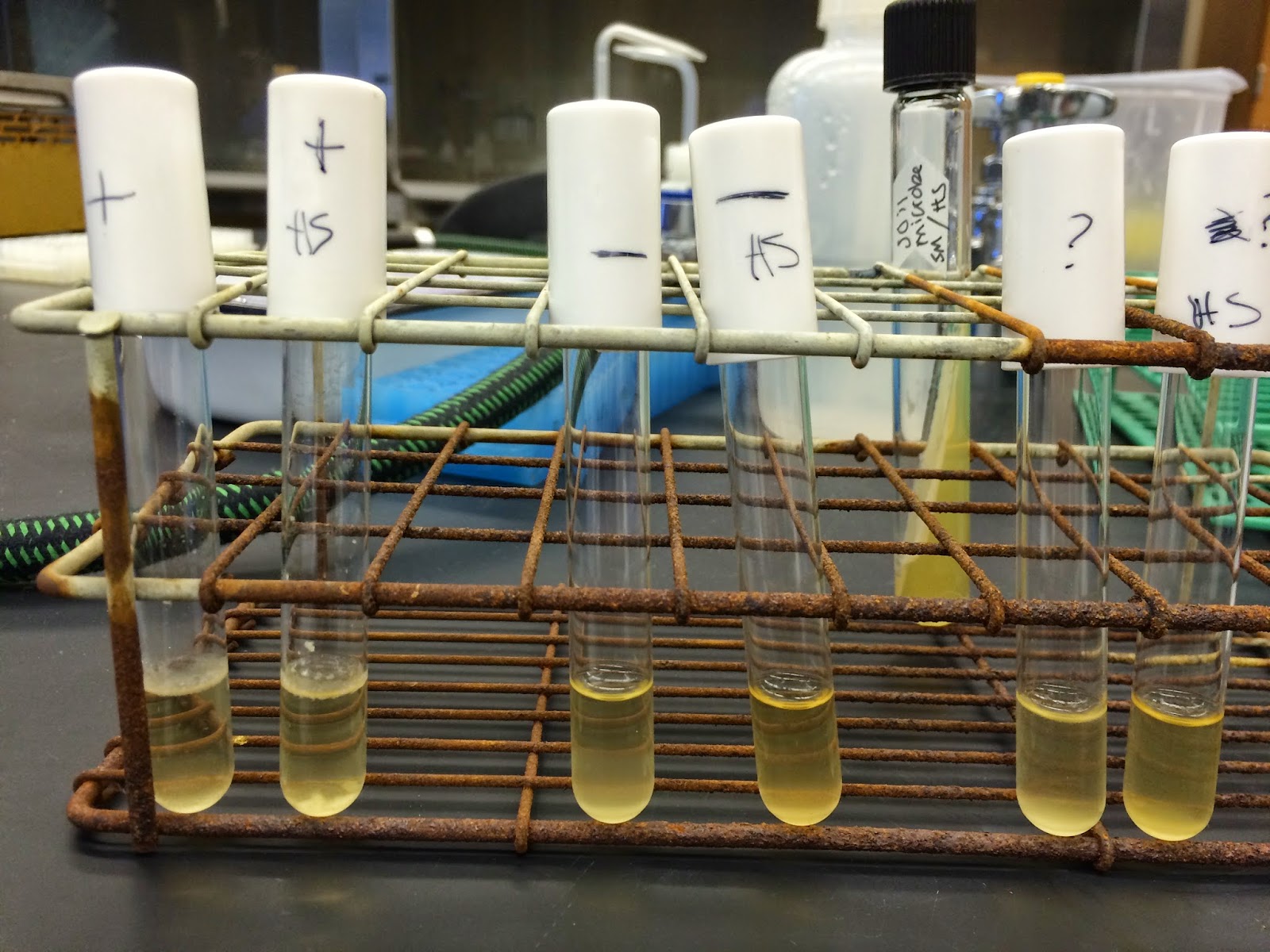
Blow is a picture that represents the results that were found
It is difficult to establish clear results from the picture, but in person it is clear that our soil sample did contain endospores. The soil sample appeared cloudy, which means that it did contain endospores. To verify this, our soil sample was compared to our positive control, Bacillus.
EDIT FROM LAB MARCH 31/2015.. (Repeated Experiment)
It was suggested that we repeat our endospore HS shock experiment to verify our results…
Blow is a picture that represents the results that were found
 |
| Repeated experiment on March 31, 2015 |
It is difficult to establish clear results from the picture, but in person it is clear that our soil sample did contain endospores. The soil sample appeared cloudy, which means that it did contain endospores. To verify this, our soil sample was compared to our positive control, Bacillus.
To further narrow down our soil microbe, a dichotomous key has been used:
Below is our journey of discovery so far….
1. Gram positive
2. Morphology, Rod Shaped (Bacilli)
3. Non-acid fast organism
4. Catalase activity
5. Endospore positive (found from the Endospore stain experiment and HS experiment)
1. Gram positive
2. Morphology, Rod Shaped (Bacilli)
3. Non-acid fast organism
4. Catalase activity
5. Endospore positive (found from the Endospore stain experiment and HS experiment)
Also, we did another experiment in lab with our soil microbe and endospore stain. The stain provided us recognition if our soil sample contained a bacterial endospore. The endospore contains a very thick wall that allows resistance to environmental conditions. We used a staining procedure called the Schaeffer-Fulton to look for endospores in bacteria. If the sample has endospores, they will appear green in pink cytoplasm.
To do this, we prepared a smear of our soil sample, a positive sample (Bacillus) and a negative control (E.coli). Each smear was heat fixed and then placed over a steaming beaker of water to heat the underside of the slide. The slides were flooded with malachite green.
Each slide was heated for five minutes. After the five minutes were up, the slides were cooled to room temperature and rinsed with water. The slides were counterstained with aqueous safranin for 30-60 seconds and rinsed with water.
To do this, we prepared a smear of our soil sample, a positive sample (Bacillus) and a negative control (E.coli). Each smear was heat fixed and then placed over a steaming beaker of water to heat the underside of the slide. The slides were flooded with malachite green.
 |
| The positive control, negative control, and soil sample smears that were prepared |
 |
| A snapshot after the malachite green was added to the slides |
 |
| Counterstain with aqueous safranin |
After they were dry, we looked at them under a microscope at 100X using oil immersion. Below are some snapshots of the slides under the microscope. The positive control and negative control showed no signs of endospores. Also, the positive control, should have contained endospores, but it was difficult to tell under the microscopes at obvious, green endospores. The microscope was a little shaky, and was difficult to see through. Our soil microbe did clearly contain endospores that were shown by in green towards the top of the slide.
 |
| Positive control (a species of Bacillus) |
 |
| Negative control (a species of E. coli) |
 |
| Our unknown soil microbe, which contained endospores shown in green (towards the top of the slide) |
Citations:
https://micro.cornell.edu/research/epulopiscium/bacterial-endospores
http://www.sciencedaily.com/articles/e/endospore.htm
http://highered.mheducation.com/sites/0072437316/student_view0/chapter27/answers_to_text_questions.html
No comments:
Post a Comment