Tuesday, April 28, 2015

Blog Post #10

Last week in lab, Hannah and I performed antibiotic sensitivity on our soil microbe in order to continue the journey of identifying our soil microbe!

Before I begin to explain our methods and results…I want to give some insight as to what antibiotics are and why they are important.  Antibiotics are powerful medicines that fight bacterial infections either by killing bacteria or keep it from reproducing. Antibiotics have no affect on viruses so they will not fight off infections such as the flu or common cold. Antibiotics are important because they can save lives and prevent infections from spreading!

Typically, if a microorganism is isolated from a patient in a hospital, they will perform antibiotic sensitivity testing on it.  The goal of this type of test is to predict the success or failure that will occur for certain types of antibiotics on that microbe. The tests are usually performed in vitro and are performed to measure the growth response of the isolated organism and its resistance or susceptibility to antibiotics.

In lab last week, we used a method called the 'disk diffusion test' to test the effectiveness of antibiotics against different species of bacteria and our unknown soil microbe.

To prepare for this procedure, we first had to start a TSB culture of our soil microbe the day before lab so that it had enough to grow.  The next day during lab we obtained four plates of antibiotic test medium.  The four plates that we chose to study were 1) unknown soil microbe, 2) E.Coli, 3) K. Pneunomonia, 4) P. Aeruginusa
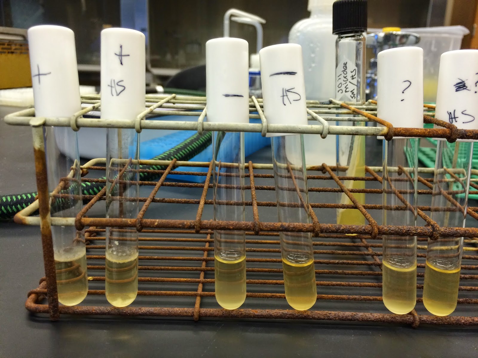
Below is a snapshot of the three different species of bacteria we used and our soil microbe before we spread them across the agar plate.

The bacteria species that we chose to test and our soil microbe

We used aseptic technique and saturated a cotton swab with each of the bacterial broth cultures that we chose to use. We spread the culture uniformly across the agar medium by wiping back and forth, covering the entire agar plate. Next, we used the antibiotic disk dispenser to place antibiotic disks uniformly on the plate.  We sterilized the forceps between each dispersion of the antibiotics. Each plate was labeled with 1,2,3,4 in a clockwise direction.  The same antibiotic would go on number 1 for all plates, a different antibiotic for number 2 on all plates, and so on. Below is a snapshot of what the plates looked like after the antibiotics were dispensed on each of the plates.
Agar plates with the antibiotics for each of the bacterial species and our unknown soil microbe plates


The plates were incubated at 37 degrees celsius for 24 hours. When we returned 24 hours later, we evaluated and analyzed each of the agar plates for resistance or sensitivity to the antibiotics.

Below is a list of the organisms that were tested and the antibiotics tested and their concentrations.

Organisms Tested:  
Unknown soil sample
E. Coli
K. Pneunomonia
P. Aeruginusa           

Antibiotics and Their Concentration:
1.) Carbenicillin (100)
2.) Erythromyclin (15)
3.) Tetracycline (30)
4.) Ampicillin (10)

Below are some snapshots of the plates…if growth occurs up to the disk, the the organism is resistant to the antibiotic. If there is a clear zone of inhibition (no growth) around the disk, then the organism is sensitive to the antibiotic.
Unknown soil microbe agar plate
There is no growth around the disk for Erythromyclin and for Tetracycline, which shows that our soil microbe is sensitive to those 2 antibiotics.  Also, there is little growth around carbenicillin, but it is unclear if the organism is resistant to it or not. There is growth all the way up to the Ampicillin disk which means that our unknown soil microbe is resistant to it.


E. Coli agar plate
The E.Coli plate shows sensitivity to both the Ampicillin, Carbenicilin, and Tetracycline antibiotics.  It is resistant to the Erythromyclin antibiotic.

K. Pneunomonia agar plate
The K. Pneunomonia plate shows antibiotic sensitivity to only the Tetracycline drug. It is resistant to Carbenicillin, Erythromyclin, and Ampicillin.

P. Aeruginusa agar plate
The P. Aeruginusa agar plate showed sensitivity to only the Carbenicillin antibiotic.  It had resistance to the Carbenicillin, Erythromyclin, and Ampicillin antibiotics. 

Our soil microbe is similar to the other bacterial species in terms that E.Coli and K. Pneunomonia was also sensitive to Tetracycline.  Also, our soil microbe, K. Pneunomonia, and P. Aeruinusa were both resistant for Carbenicillin and Ampicillin.

In a previous study, we identified our soil microbe to be Gram positive. 


So far in the dichotomous key we have identified our soil microbe:
1. Gram positive
2. Morphology, Rod Shaped (Bacilli)
3. Non-acid fast organism
4. Catalase positive
5. Endospore positive
6. Motile
7. Nitrate reduction
8. Alpha hemolysis
The dichotomous key indicates that our soil microbe belongs to the genus Streptococcus!

In order to further identify our soil microbe to the particular Streptococcus species, we need to do a P/A disc test. The P disc test is an optochin susceptibility test and the A disc test is a bacitracin susceptibility test. To do this, we would need to do an experiment very similar to the one we performed in this week's lab to test antibiotic resistance/sensitivity. However, we would need to inoculate a disc that contained optochin in the center of the agar surface. The plate would need to be stored for 24 hours at 35-37 C. If the growth went all the way to the margin of the disc, then it is resistant to the chemical. If sensitive, the species that would be identified is Streptococcus pneumonia. If resistant, the species would be Streptococcus mitts.
The bacitracin test would be the same method used for the optochin test; however, the disc would contain bacitracin. If sensitive, the species that would be identified is Streptococcus progenes. If resistant, the species identified would be Streptococcus galactiae. 

References:
http://www.uphs.upenn.edu/bugdrug/antibiotic_manual/amt.html

http://www.vumicro.com/vumie/help/VUMICRO/Optochin_Susceptibility.htm

http://www.vumicro.com/vumie/help/VUMICRO/Bacitracin_Susceptibility.htm

Monday, April 20, 2015

Blog Post #9


blood agar plates

Last week in lab Sammy and I tested our mystery microbe's hemolytic abilities. We did this by using blood agar plates containing general nutrients and 5% sheep blood. We used the T-streak method to inoculate S. aureus, S. epiderdimis, and our unknown microbe onto three separate plates. We then incubated the plates at 37 degrees Celcius and checked them at 24 and 48 hours to check for hemolysis. 

24 hours: left: unknown, top right: S. aureus,
bottom right: S. epididermis


Unknown after 24 hours of incubation


According to our lab handout there can be divided into three groups – alpha, beta, and gamma – based upon their hemolytic activity on blood agar. Beta (β) hemolysis is defined as complete or true lysis of red blood cells. Alpha (α) hemolysis is the reduction of the red blood cell hemoglobin to methemoglobin in the medium surrounding the colony. Gamma (γ) hemolysis indicates the lack of hemolysis. After 24 hours there is already significant growth on some of our plates. Our mystery microbe grew very quickly on the blood agar and is seems to fit the description of an alpha hemolytic bacteria. As you can see in the photos there is a clear dark discoloration surrounding the colonies on the plate.


Unknown after 48 hours
Plates after 48 hours


Bacteria are able to lyse red blood cells by producing enzymes called homolysins. Thers enzyemes function by destroying the outer membrane of a red blood cell. Some create pores in the membrane, weakening it, while other hydrolyze the phospholipid components of the cell's phospholipid bilayer. Hemolytic microbes are more virulent than non-hemolytic microbes because they destory iron-containing red blood cells that fight infection. This results in a reduced number of red blood cells in the body and therefore reduced oxygen transportation to the body which can be dangerous. I would not expect a typical soil microbe to be capable of hemolysis because most of them are not pathogenic. 

We've been attempting to identify our mystery microbe this semester by using a dichotomous key. This week we determined that our microbe is capable of alpha hemolysis.


1. Gram positive
2. Morphology, Rod Shaped (Bacilli)
3. Non-acid fast organism
4. Catalase positive 
5. Endospore positive
6. Motile
7. Nitrate reduction
8. Alpha hemolysis

The dichotomous key indicates that our microbe belongs to the genus Streptococcus! 

http://en.wikipedia.org/wiki/Hemolysin
http://en.wikipedia.org/wiki/Streptococcus

Monday, April 13, 2015

Blog Post #8

Last week in lab, Hannah and I did an experiment to determine if our soil microbe reduced nitrate or not.  Nitrogen is a basic element of life and is a component of both proteins and nucleic acids.  Nitrogen is very abundant in the atmosphere; however, it is not in the accessible for that living organisms need it to be in.

In order to test if our soil microbe was a nitrate reducer or not, we used a positive control (P. Auroginosa), a negative control (E. Coli), and our soil microbe and inoculating 3 cultures (1 of each).  Each of the samples were incubated for 24-48 hours.  Our soil microbe is a fast grower, so we returned 24 hours later.

A snapshot before we returned to lab 24 hours later

The second day, we returned to observe the Durham tube in each sample.  If there were bubbles in the Durham tube, then the organism was not a fermenter.  However, our sample did not have any bubbles present so we added 8 drops of reagent A and 8 drops of reagent B and mixed well.  Once the drops were added, the sample turned red.  This means that the microbe reduced nitrate to nitrite!!  Below are snapshots of our Durham tubes during the experiment.

After reagent A & B were added, our sample turned red

Reagent A & B that were added to our soil sample
In order to further understand nitrate reduction, let me explain some information from a broader, biological context.  Most bacteria use the cellular structures called flagella for motility.  They attach to the cell surface and provide a 'swimming' movement.  There are also flagellar arrangements for bacteria that can be determined by staining and microscopic observation.  

Nitrate reduction indicates that the organism can use NO3- as an electron acceptor.  Also, nitrite may be reduced to either NO, N20, N2, and NH3 depending on the enzyme system of the organism and the atmosphere it is growing in.  Nitrate reduction often involves a shift to anaerobic metabolism.  Nitrate reduction plays a key role in the nitrogen cycle.  I think that some microbes evolved to be nitrate reducers, because nitrate reduction is one of the most fundamental biological processes that accounts for tons of inorganic nitrogen.  Simply stated, since nitrogen is a basic element for life, nitrogen reductase evolved in order to maintain the nitrogen cycle, which allows biological processes to be carried out successfully. 

I think that some microbes did not evolve to be nitrate reducers, because nitrogen was in a form that was accessible and usable to that particular organism.  Organisms that did not evolve to be nitrate reducers were able to use nitrogen in the form that it was already in, rather then converting it to a different form. 

Along our journey to identify our soil microbe, we have been keeping track by using a dichotomous key.  Below is the identification of our mysterious soil microbe thus far:

1. Gram positive
2. Morphology, Rod Shaped (Bacilli)
3. Non-acid fast organism
4. Catalase activity
5. Endospore positive
6. Motile
7. Nitrate reduction

Citations:

http://www.nature.com/scitable/knowledge/library/the-nitrogen-cycle-processes-players-and-human-15644632

http://www.microbelibrary.org/library/laboratory-test/3660-nitrate-and-nitrite-reduction-test-protocols


Monday, April 6, 2015

Blog Post #7

This past week in lab Sammy and I tested the motility of our microbe. Motility is the ability of a microbe to move on its own. By using a special, soft agar we can see motile bacteria grow throughout the medium and non-motile bacteria grow only where the inoculating needle stabbed the agar. We tested a positive control (E. coli) and a negative control (S. aureus) in the agar to compare to our mystery microbe. Unfortunately, this specific E. coli we used was actually not motile so both our controls were negative. Our mystery microbe ended up not growing within the agar or along the line of inoculation but instead on the surface of the agar. Since our triple sugar test a few weeks ago showed that our microbe can tolerate anaerobic growth it is most likely that our microbe is motile and it “swam” out of the stab and to the surface of the agar.


Bacteria need to possess flagella, threadlike organelles extending from the cells outer membrane, to be capable of motility. Different bacteria vary in their number of flagella from one to numerous. Bacteria can also move using other cellular structures including the lesser-understood gilding motility, which does not involve flagella. Another bacterial group called spirochetes move by using axial filaments, which are similar to flagella.

 
example of flagellum and axial filament structures
http://ncse.com/book/export/html/890 http://classes.midlandstech.edu/carterp/courses/bio225/chap04/lecture3.htm
Some microbes have evolved to be motile when it is evolutionarily beneficial in their environment. Some microbes need to be able to flee from a harmful environment and towards a desired environment. This type of movement is described in our textbook as chemotaxis. Although it seems evolutionarily beneficial to be motile some bacteria cannot move on their own. A possible explanation for this is that these bacteria can survive in harsher environments than motile bacteria and therefor do not need the energetically costly cellular structures necessary for motility.

So far the dichotomous key has shown us that are microbe fits the following criteria..
1. Gram positive
2. Morphology, Rod Shaped (Bacilli)
3. Non-acid fast organism
4. Catalase activity
5. Endospore positive 
The link for the key we have been using doesn't seem to be working currently but the next step, I believe, was whether our microbe was motile or non motile. According to our soft agar test we can tentatively say our microbe is motile!
6. Motile

Citations: 


Sunday, March 29, 2015

Blog post #6

Last week in lab, Hannah and I verified that our soil microbe did produce endospores.  First, it is important to understand what an endospore is.  An endospore is a dormant, tough, non-reproductive structure produced by a small number of bacteria.

The function of an endospore is to ensure the survival of a bacterium through periods of environmental stress.

The benefits of an endospore is that they protect the bacteria's genome and a small amount of cytoplasm.  Endospores offer an advantage to bacteria that are able to produce them because it protects them from being destroyed.  Bacteria that does have the ability to produce endospores will have a greater chance of survival.  Bacteria that is not able to produce endospores could easily die off from harsh environmental conditions.

Some microbes have evolved to form endospores because microbes sense and adapt to changes in the environment.  They also might have evolved endospores due to response for nutrient deprivation.  Also, some microbes might not have evolved to form endospores because microbes are already adapted to conditions that are favorable for survival.  Some of these stresses could include high temperature, high UV radiation, chemical damage, etc.  A microbe may not evolve the formation of endospore because it already has nutrients that are needed and is adapted to changes in the environment.

In lab, we used aseptic technique to inoculate six 2 mL tubes of tryptic soy broth (TSB) with bacteria.  The specific cultures that we made were:
1. 2 tubes with positive Bacillus control
2. 2 tubes with negative E.coli control
3. 2 tubes with unknown soil sample
For each of the bacterium's, we labeled one tube 'HS' for heat shock.

3 tubes that contain the positive, negative, and soil microbe

Six tubes that we made using the positive control, negative control, and soil microbe before the 3 heat shocked were placed inside the water bath
After we transferred the HS tube of each bacterium, the three tubes were transferred to the 80 degree water bath.  The tubes were incubated in the water bath for 10 minutes.
Our 3 samples that were placed in the 80 degree water bath (HS samples)

After the 10 minutes, the samples were removed and all the tubes (6 total) were left on the bench to grow.

Below are some snapshots of the observations from our samples:
After 48 hours, the tubes were vortexes and observations were recorded

To interpret our results, it is important to not that all of our HS samples did have growth, which was observed by cloudiness in the tubes.  Since the E-coli served as the negative control, and did not contain endospores, the HS sample was clear because it did not survive the water bath, which is seen from the picture above.

The positive control, Bacillus served as the control that contained endospores.  This means that HS sample did survive the water bath, and thus appears cloudy.

For our soil microbe, the HS sample is exactly like the E-coli negative control.  The HS sample is clear, which means that no endospore formation was present and the soil sample did not survive the high temperatures from the water bath.

EDIT FROM LAB MARCH 31/2015.. (Repeated Experiment)
It was suggested that we repeat our endospore HS shock experiment to verify our results…
Blow is a picture that represents the results that were found
Repeated experiment on March 31, 2015

It is difficult to establish clear results from the picture, but in person it is clear that our soil sample did contain endospores.  The soil sample appeared cloudy, which means that it did contain endospores.  To verify this, our soil sample was compared to our positive control, Bacillus.


To further narrow down our soil microbe, a dichotomous key has been used: 
Below is our journey of discovery so far….
1. Gram positive
2. Morphology, Rod Shaped (Bacilli)
3. Non-acid fast organism
4. Catalase activity
5. Endospore positive (found from the Endospore stain experiment and HS experiment)


Also, we did another experiment in lab with our soil microbe and endospore stain. The stain provided us recognition if our soil sample contained a bacterial endospore.  The endospore contains a very thick wall that allows resistance to environmental conditions. We used a staining procedure called the Schaeffer-Fulton to look for endospores in bacteria.  If the sample has endospores, they will appear green in pink cytoplasm.

To do this, we prepared a smear of our soil sample, a positive sample (Bacillus) and a negative control (E.coli).   Each smear was heat fixed and then placed over a steaming beaker of water to heat the underside of the slide.  The slides were flooded with malachite green.
The positive control, negative control, and soil sample smears that were prepared

A snapshot after the malachite green was added to the slides
Each slide was heated for five minutes.  After the five minutes were up, the slides were cooled to room temperature and rinsed with water.  The slides were counterstained with aqueous safranin for 30-60 seconds and rinsed with water.  
Counterstain with aqueous safranin


After they were dry, we looked at them under a microscope at 100X using oil immersion.  Below are some snapshots of the slides under the microscope.  The positive control and negative control showed no signs of endospores.  Also, the positive control, should have contained endospores, but it was difficult to tell under the microscopes at obvious, green endospores.  The microscope was a little shaky, and was difficult to see through.  Our soil microbe did clearly contain endospores that were shown by in green towards the top of the slide.
Positive control (a species of Bacillus)

Negative control (a species of E. coli)

Our unknown soil microbe, which contained endospores shown in green (towards the top of the slide)


Citations:
https://micro.cornell.edu/research/epulopiscium/bacterial-endospores

http://www.sciencedaily.com/articles/e/endospore.htm

http://highered.mheducation.com/sites/0072437316/student_view0/chapter27/answers_to_text_questions.html

Monday, March 23, 2015

Blog Post #5

In our most recent lab meeting Sammy and I tested our mystery microbe for catalase activity. To accomplish this we first tested some controls that we could compare our microbe to. We used a positive (S. epideidermis) control and a negative (E. faecalis) control.  We observed bubble formation with our positive control when a drop of 3% H2O2 was added and no reaction with our negative control.


We then added the drop to our microbe and observed bubble formation indicating a positive result for our catalase test. A potential explanation for the evolvement of catalase activity in certain microbe is the aerobic microbes adapting to fight against elements that anaerobic microbes do not encounter such as oxygen. 


The other experiment we did regarding our microbe was the triple sugar iron test to determine carbohydrate metabolism. The TSI slant provides the culture with an aerobic and anaerobic environment in which to grow. This allows us to observe how different bacterium ferment and metabolize. All five of our control tubes grew as expected. I compared the information provided to us about the predicted reactions for each bacterium and confirmed the results by observing the color of the TSI agar of each control after incubation.


E. coli

P. vulgaris

P. aeruginosa
B. Megaterium 


For our unknown microbe, the media at the top of the tube was pink/red indicating an alkaline reaction. The bottom, or butt ,of the tube was yellow indicating an acidic reaction. There was no evidence of hydrogen sulfide formation. These results mean that our microbe metabolized glucose 
but not sucrose or lactose.


In the last blog post, Sammy followed the dichotomous key up until the question regarding catalase activity…
Dichotomous Key
Below, I have used a dichotomous key in order to begin identifying our microbe…Below is the steps that I followed in the dichotomous key:
1. Gram Positive
2. Morphology, Rod Shaped (Bacilli)
3. Non-acid fast organism

Now we know that our microbe is catalase (+) so we can continue!
4. Catalase positive


We know that our microbe metabolized glucose in both the aerobic and anaerobic areas of the slant so I’m unsure of the answer to the next step in the key. We might have to have further discussions to determine the answer.