Monday, March 9, 2015

Blog Post #4

Last week in lab, Hannah and I performed an acid stain test using two different stain samples:
1) a non-acid fast control (M. Luteus)
2) soil microbe

M. Luteus (non-acid fast control)

Soil Microbe
One day prior to lab, we prepared a pure culture of our soil microbe on a TSA slant.

During this acid stain procedure, we used an aseptic technique and smeared a sample of our soil microbe and the control culture onto a slide, along with a loopful of water.  We mixed the bacteria with the water and allowed it to dry. Then, we put our slide through the bunsen burner and placed the slides over a beaker with steaming water.  The slides were then flooded with carbol-fushin and cooled/rinsed with water.  The slides were depolarized with acid-alcohol until the color stopped coming off.  Lastly, the slide was flooded with methylene.

Below is a picture of the control slide & soil microbe over steaming water:

We then observed our acid fast stain slides under the microscope at 100X magnification. Sidenote:  (Oil immersion was added to the slide).

Below is a picture of the control and our soil microbe under the microscope:
The soil microbe had a blue tint with many blue rods (bacilli) distributed across the slide.
Soil Microbe
It was observed under the microscope to have very small, round, blue circles distributed evenly across the slide.
M. Luteus
Acid-Fast Stain & Background:
It is important to include some background information about an acid fast stain and what it actually is to further understand and interpret our results.  An acid fast stain is a stain that is used to identify organisms, such as members of the genus, Mycobacterium. Acid fast organisms usually have waxy cell walls that are impermeable and contain many lipids and fatty acids.  Usually to get through this cell wall, heat is involved.

It was found that our soil microbe, was non-acid fast, which means that it does not have a waxy cell wall.  It was also gram (+) in terms that a peptidoglycan wall is present. 

Dichotomous Key
Below, I have used a dichotomous key in order to begin identifying our microbe…Below is the steps that I followed in the dichotomous key:
1. Gram Positive
2. Morphology, Rod Shaped (Bacilli)
3. Non-acid fast organism
4. …The next question asks if the organism is catalase (+) or catalase (-)…Unfortunately, at this time in the experiment we do not know the answer to this question yet so we will have to look forward to next week's lab in order to further identify our mysterious microbe!


Monday, March 2, 2015

Post #3


During lab this week we Gram stained several different bacterium. First we made two control stains one Gram positive, B. megaterium, and one Gram negative, K. pneumoniae.  After viewing them under the microscope the Gram positive sample is clearly purple and the Gram negative sample is pink.

Gram positive control
Gram negative control

 We then Gram stained our mystery microbe to determine what category it fell into. When viewed under the microscope our mystery microbe was purple indicating that it is Gram positive. It also appeared rod shaped or Bacillus.


soil microbe
soil microbe under microscope

 After checking out our microbe under the microscope we streaked our soil microbe on a MacConkey agar plate along with B. megaterium, K. pneumoniae, and P. aeruginosa. We also streaked these bacterium on a Rose-Bengal plate to determine if their was any fungi present in our sample.


Our MacConkey agar confirmed our Gram staining. B. megaterium and our mystery microbe both did not grow on the agar indicating that they’re Gram positive. K. pneumoniae and P. aeruginosa successfully grew on the plate demonstrating that they are Gram negative. Also, there was no growth for our microbe on the Rose-Bengal plate. 


There are several differences between the cell wall structure of Gram positive and Gram negative bacteria. Gram positive bacteria have a cytoplasmic membrane with a thick peptidoglycan outer layer. Gram negative bacteria has a thin peptidoglycan layer sandwiched between two cytoplasmic membranes.  The outer membrane of the Gram negative  bacteria contains phospholipids, proteins, and lipopolysaccharide (LPS). LPS is an endotoxin that contains Lipid A.  When the Gram negative bacteria is killed the endotoxin is released causing negative effects in the host. This feature of the cell wall can cause issues when treating Gram negative bacterial infections. The cell wall structure also is resistant to many antibiotics making the infections difficult to treat. 

Monday, February 23, 2015

Blog Post #2

Last week, Hannah and I took a soil sample from the Ginkgo tree in front of the Stevens Science building and used plates to dilute microorganisms that could be present in the soil.  The plates ranged from 10^-3, 10^-4…10^-7.  Below are some snapshots of the colonies of microorganisms that were present on the plates.

Biodiversity is a key part when identifying microbes.  As you can tell from our pictures below, there was a wide scale of biodiversity among the microbes that were present in each of our dilution plates.  In  the 10^-3 plate, there were too many colonies to count, but the colonies were small and scattered randomly across the plate.  Towards the left of the plate you can see a large colony that is smooth and 'gel like'.
10^-3
In the 10^-4 plate there are 28 colonies present and most of these colonies are 'slime-like' and have a sticky appeal to them.  They are much larger then the colonies present on the 10^-3 plate.
10^-4
Biodiversity here is indicated by 8 colonies are different size.  Most are circular in structure and also have a glob-like look to them.  The smaller colonies appear to be smooth and are not as 'gel like'.  Also, it is important to observe a white fluffy colony that is attached to a colony on the plate.  This is a fungi colony that has grown on the plate.
10^-5
There are only 2 colonies present on this plate.  One colony is smaller in shape and has a white fuzzy look it to.  Could it be a fungi, perhaps?
10^-6
Lastly, in our 10^7 plate, there is no colonies present due to the dilution.
10^-7
In the rose bengal agar, there was one colony of fungi that had grown.  It was white and fungi in shape, yet quite small in comparison to the bacteria that was present on the other plates.
10^-4 (Fungi)

** It is very important to note and observe that bacteria outnumbers the fungi**


In lab, we counted the number of colonies that were present on each plate.  To do this, we divided the number of colonies of the volume of the sample spread times the dilution factor for each plate.  Below is a list of the data that was obtained after calculations:

10^-3   (TMTC)………….<280,000 microbes/1 g soil
10^-4   (28)……...………280,000 microbes/1 g soil
10^-5   (8)…….………….800000 microbes/1 g soil
10^-6   (2)…….………….2000000 microbes/1 g soil
10^-7   (0)…….………….0 microbes/1 g soil


Why does Biodiversity matter?

There are many ways to answer this question, because biodiversity is very important not just in terms of science, but to human application as well…

1) benefits that biodiversity provides us (such as drinking water, food, air)
2) medicinal resources
3) agricultural resources
4) wood products
5) diversity in genes
6) recreational activities- such as hiking, tourism, etc…pure enjoyment of nature!

Without biodiversity, we would not be able to survive and thrive as human beings.  Biodiversity is often overlooked, but it is important to preserve biodiversity so that animals, plants, and microorganisms won't go extinct and we can continue to live on this earth as a human species!

Citation: http://www.globalissues.org/article/170/why-is-biodiversity-important-who-cares

Last week in lab, our goal was to establish a pure culture of soil microbes using the T streak strategy from the soil sample plates from the previous week.

Hannah used a colony from the 10^-4 plate.  Below is a picture of the colony that Hannah took a sample from.  It is the colony towards the bottom of the plate that is medium sized, and perfectly circular.

24 hours later, the plate was observed and below is a picture of the results that were obtained:

I (Samantha), chose a colony from the 10^-5 plate that was a larger colony, to see the difference that it would have after the 24 hours.  Below is a picture of the colony that was swabbed.
After 24 hours, the plate was observed and the picture below shows the results front the T streak application.  It was observed that the pure culture of soil microbe was present after the second quadrant of the T streak.


Monday, February 16, 2015

Blog Post #1


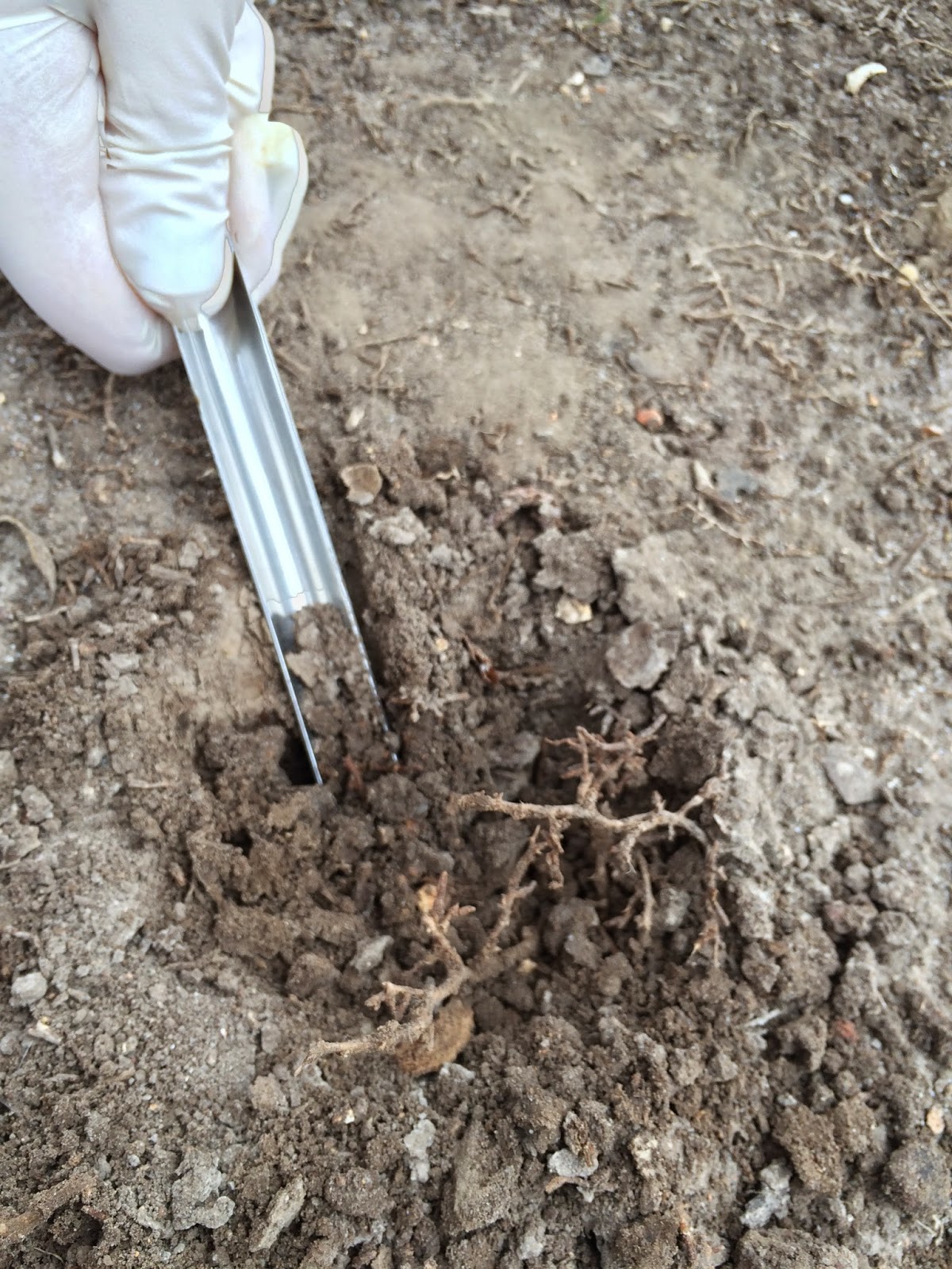
Sammy and I decided to collect soil near the Ginkgo tree next to the Humanities building. On the day we collected the habitat was cool and dry. However, on a sunnier day the soil could definitely experience direct sunlight.  The Ginkgo tree is fairly isolated from other plants. Two sidewalks separate it from the bushes along the sides of Humanities and Munger.


The top layer of soil was very dry but when we dug a few inches deeper into the ground the soil was visibly moister. There were definitely rhizospheres in the soil as we collected. Before digging, we put on latex gloves and wiped down our trowel and scoopula and laid them on clean paper towels. After digging into the soil with the trowel we used the scoopula to collect 15ml of soil. Once we had our soil we headed back into the lab and weighed out 0.5g or our sample and mixed it with 50ml of sterile water. We then set this mixture aside and allowed the larger soil particles to settle at the bottom of the tube.

While the soil in out tube settled we labeled five sterile microfuge tubes and five tryptic soy agar plates with 10^-3 through 10^-7. We then pipetted 0.9ml of sterile water into each microfuge tube. Once most of the soil settled to the bottom of the tube we pipetted 1ml of the mixture into the microfuge tube labeled 10^-3 and mixed well. We then transferred 0.1ml from the 10^-3 microfuge tube to the tube labeled 10^-4 and mixed well. We continued this serial dilution ending with the tube labeled 10^-7.

After all the dilutions were prepared we transferred 0.1ml of each dilution on to the corresponding agar plate and spread the cells evenly across the plate.  Lastly, we spread 0.1ml of the 10^-4 dilution onto a plate of Rose-Bengal agar plate. We then inverted the plates and left them to incubate at room temperature for about a week. After 6 days of incubating on the lab counter our plates are definitely showing some growth! Our plate with the 10^-3 dilution was growing so much that Dr. Hanson had to put in the fridge to slow things down.   

Agar plates before incubation (2/10/15) 
Agar plates after incubation (2/16/15)